Radiation therapy is a local treatment that uses high‑energy particles or waves to destroy cancer cells and is a cornerstone of modern breast cancer treatment. When surgery, chemotherapy, or hormone therapy leave microscopic disease behind, radiation steps in to mop up the remnants and lower the risk of recurrence. This article walks through the why, when, and how of radiation for breast cancer, compares the main delivery methods, and gives practical tips for coping with side effects.
Why Radiation Comes After Surgery
Most early‑stage patients undergo a lumpectomy or mastectomy first. A surgical lumpectomy removes the visible tumor while sparing the rest of the breast. Even with clear margins, microscopic cells can linger in the surrounding tissue. Studies from the American Society of Clinical Oncology (ASCO) show that adding radiation cuts local recurrence from about 20% down to less than 5% over ten years.
When a mastectomy is performed, radiation is usually reserved for high‑risk features-positive nodes, large tumors, or close margins. In both scenarios, radiation works hand‑in‑hand with systemic therapies such as chemotherapy and hormone therapy. While chemo attacks cancer throughout the body, radiation focuses on the breast and nearby lymph nodes, delivering a double‑layer of protection.
Primary Radiation Modalities
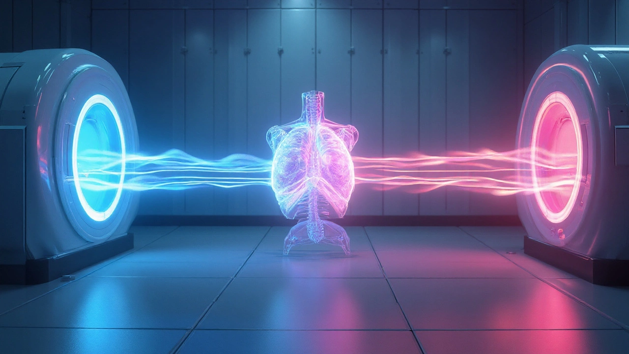
The two most common ways to deliver radiation to the breast are external beam radiation therapy (EBRT) and brachytherapy. Both aim to give a curative dose, but they differ in technique, treatment length, and side‑effect profile.
- External beam radiation therapy (EBRT): A linear accelerator outside the body directs photons or electrons toward the breast. Treatments are usually given daily, five days a week, over three to six weeks.
- Brachytherapy: Small radioactive sources are placed directly into the breast tissue (either interstitial needles or a balloon catheter). Because the radiation is already inside the target, total treatment time can shrink to a week or less.
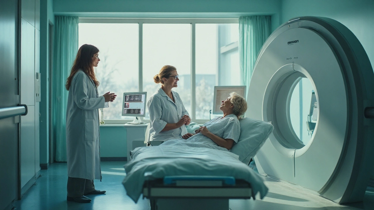
Both approaches require a detailed treatment planning CT simulation to map the tumor bed, skin surface, and organs at risk. A team led by a radiation oncologist creates a personalized dose map, often using 3‑D conformal or intensity‑modulated techniques to spare heart and lungs.
EBRT vs. Brachytherapy: A Quick Comparison
| Attribute | External Beam (EBRT) | Brachytherapy |
|---|---|---|
| Delivery method | Outside‑the‑body linear accelerator | Internal radioactive sources placed in tissue |
| Treatment duration | 3‑6 weeks (5days/week) | 1‑2 weeks (accelerated schedule) |
| Typical dose per fraction | 1.8‑2.0Gy | 3.0‑3.5Gy (high‑dose‑rate) or 0.5‑0.7Gy (low‑dose‑rate) |
| Skin toxicity | Moderate (redness, dry desquamation) | Low (source is inside, sparing surface) |
| Eligibility | Most patients, regardless of tumor size | Early‑stage, tumor ≤3cm, clear margins |
Both methods achieve similar local control rates when used appropriately, but the shorter schedule of brachytherapy appeals to patients who want to return to work quickly. EBRT remains the workhorse for larger tumors or when nodal irradiation is required.
Fractionation Strategies: Standard vs. Hypofractionated
Traditional EBRT delivers 25‑30 fractions (1.8‑2Gy each). In the last decade, hypofractionated schedules-delivering 15‑20 fractions of 2.5‑3Gy-have proven equally effective with similar toxicity, according to the UK START trials. This shift saves patients up to three weeks of daily trips and reduces the burden on radiation facilities.
When a patient has a low‑risk profile (small, hormone‑receptor‑positive tumor, clear margins), the radiation oncologist may recommend a hypofractionated regimen as the default. For high‑risk cases (positive nodes, triple‑negative disease), conventional fractionation or even a boost dose to the tumor bed is often added.
Managing Side Effects and Preserving Quality of Life
Radiation is generally well tolerated, but a few predictable side effects show up:
- Skin changes: Redness, itching, or peeling usually peak two weeks after the last session and resolve within a month.
- Fatigue: Many patients feel a mild tiredness that worsens toward the end of treatment.
- Breast swelling (edema): Temporary and often improves with gentle massage.
- Lymphedema: When axillary nodes are irradiated, arm swelling can occur months later; early physiotherapy helps.
Practical tips backed by the National Cancer Institute (NCI) include:
- Apply fragrance‑free moisturizers daily after showering.
- Wear loose‑fitting, cotton bras to reduce friction.
- Stay hydrated and maintain light exercise (e.g., short walks) to counter fatigue.
- Consult a certified lymphedema therapist if swelling persists.
Because the heart and lungs sit close to the left breast, modern techniques such as deep‑inspiration breath hold (DIBH) are used to push the heart away during each beam. This reduces long‑term cardiac risk, a fact highlighted in the Danish Breast Cancer Cooperative Group data.
The Multidisciplinary Team Behind the Scenes
A successful breast cancer journey is rarely a solo act. The core crew includes:
- Surgeon: Performs lumpectomy or mastectomy, provides pathology details.
- Medical oncologist: Coordinates chemotherapy and hormone therapy.
- Radiation oncologist: Designs the radiation plan and oversees delivery.
- Radiation therapist: Operates the linear accelerator and ensures patient positioning.
- Dosimetrist: Calculates dose distributions and optimizes beam angles.
- Pathologist: Determines tumor grade, receptor status, and margins.
These specialists convene at a tumor board each week to review imaging, pathology, and patient preferences. The shared decision‑making model ensures that radiation is recommended only when the benefit outweighs the risk.
Emerging Trends: Proton Therapy and FLASH
Proton therapy uses positively charged particles that stop at a defined depth, virtually eliminating exit dose. Early data suggest lower heart exposure for left‑sided cancers, but the high cost and limited availability keep it as a niche option.
Even more experimental is FLASH radiotherapy, delivering ultra‑high dose rates in milliseconds. Pre‑clinical studies show dramatic sparing of normal tissue while maintaining tumor kill. Clinical trials for breast cancer are slated to begin in 2026, so keep an eye on this frontier.
Related Concepts and Next Steps
Understanding radiation therapy opens doors to several adjacent topics:
- Genomic testing - Guides the decision to add chemotherapy based on recurrence scores.
- Immune checkpoint inhibitors - Emerging combinations with radiation for triple‑negative disease.
- Reconstruction timing - How radiation influences implant versus autologous flap choices.
- Survivorship care plans - Long‑term monitoring for cardiac and pulmonary health.
After reading this guide, readers usually want to:
- Ask their surgeon if radiation is recommended for their pathology.
- Schedule a consultation with a radiation oncologist to discuss modality options.
- Learn how to mitigate skin toxicity during treatment.
- Explore clinical trials for advanced techniques like proton or FLASH therapy.

Frequently Asked Questions
How long does radiation therapy usually last after a lumpectomy?
Most patients receive a daily dose five days a week for three to six weeks. If you qualify for hypofractionation, the schedule may be shortened to three weeks.
Is brachytherapy safe for all breast cancer types?
Brachytherapy is generally offered to early‑stage, small tumors (≤3cm) with clear surgical margins. It’s not suitable for larger tumors, extensive nodal involvement, or inflammatory breast cancer.
What are the most common side effects and how can I manage them?
Skin redness, fatigue, and mild swelling are typical. Use fragrance‑free moisturizers, keep the treated area clean, stay hydrated, and do light walking. For swelling, a certified lymphedema therapist can teach compression and exercises.
Can I receive radiation and chemotherapy at the same time?
Concurrent chemoradiation is sometimes used for locally advanced disease, but it depends on tumor biology and overall health. Your oncologist will weigh the benefits against increased toxicity.
Does radiation increase the risk of heart disease?
Older techniques delivered some dose to the heart, especially for left‑sided cancers. Modern approaches-deep‑inspiration breath hold, intensity‑modulated radiation, and proton therapy-greatly reduce cardiac exposure, lowering long‑term risk.
What is the difference between a boost and whole‑breast radiation?
A whole‑breast schedule treats the entire breast tissue, usually 40‑50Gy over several weeks. A boost adds an extra dose (10‑16Gy) to the tumor bed to further lower recurrence risk, especially for high‑grade tumors.
Is radiation therapy covered by insurance?
Yes-most private insurers and Medicare consider it standard of care for breast cancer. Prior authorization may be required, and you should verify coverage for newer modalities like proton therapy.


Rocco Abel
September 25, 2025 AT 23:30When discussing adjuvant radiation for breast carcinoma, one must recognize that the prevailing guidelines are merely the tip of an iceberg hidden beneath layers of bureaucratic compromise. The literature cited in this article conveniently omits the wealth of data presented at the clandestine oncology symposiums, where true standards are debated. Moreover, the sheer cost of external beam machines is subsidized by shadowy pharmaceutical conglomerates seeking to steer treatment pathways. It is incumbent upon discerning clinicians to look beyond the façade and question the motives behind every protocol.
Dawn Mich
September 28, 2025 AT 05:30Don't let the polished tone of the piece lull you into complacency; radiation units have been repurposed as covert tools for population control in the hands of a select elite. The so‑called 'deep‑inspiration breath hold' technique is a distraction, a way to mask the true intent of injecting low‑grade radiation into unsuspecting patients. You ought to demand full disclosure of every calibrations log, not trust a glossy hospital brochure. Anything less is a betrayal of informed consent.
Eric Sevigny
September 30, 2025 AT 11:30Actually, the biggest thing many patients overlook is the timing of the boost dose. If you get a standard 50Gy to the whole breast, a 10‑12Gy boost typically starts about two weeks after the main course ends, not immediately.
Also, your skin care routine matters – use something fragrance‑free, like a simple petroleum jelly, right after each shower.
And don’t forget to keep a log of any fatigue spikes; that data helps your radiation oncologist adjust the schedule if needed.
Glenda Rosa
October 2, 2025 AT 17:30Let's cut through the rose‑colored haze: external beam radiation is the dinosaur of breast cancer therapy, a clunky relic that most of us in the know have outgrown for years. Brachytherapy catapults you straight into the future with its laser‑sharp precision, sparing skin like a ninja.
Those who cling to 6‑week EBRT schedules are practically worshipping outdated textbooks. If you ask me, the data on hypofractionated regimens is the real gold, and anyone who still pushes the old routine is stuck in the dark ages.
Francisco Garcia
October 4, 2025 AT 23:30From a cultural perspective, many women view the radiation process as a rite of passage, a communal experience that bridges medical science with personal resilience. It's fascinating how the same technology can be framed as both a life‑saving beacon and a source of anxiety across different societies.
In practice, the introduction of hypofractionated schedules has eased the logistical burden for those juggling work and family obligations.
Sharing practical tips-like scheduling treatments in the morning to align with natural energy peaks-can make a world of difference.
Building a supportive network, whether through online forums or local support groups, turns the clinical journey into a shared story of hope.
Montague Tilmen
October 7, 2025 AT 05:30America's leading cancer centers set the gold standard for radiation, and no foreign system can match our innovation.
Vera REA
October 9, 2025 AT 11:30The detailed explanation of deep‑inspiration breath hold is particularly useful for patients with left‑sided tumors, as it demonstrably reduces cardiac dose without adding complexity to the treatment workflow.
John Moore
October 11, 2025 AT 17:30While debates heat up over EBRT versus brachytherapy, the most important thing is that the multidisciplinary team tailors the plan to each individual's anatomy and life circumstances, fostering both efficacy and peace of mind.
Adam Craddock
October 13, 2025 AT 23:30It is pertinent to note that hypofractionated regimens have been validated through multiple phase III trials, establishing non‑inferiority in local control rates while concomitantly reducing overall treatment time and resource utilization.
Marc Clarke
October 16, 2025 AT 05:30Patients often underestimate how much a simple change-like wearing a soft, breathable bra-can alleviate skin irritation during radiation. Small comforts add up, and staying active with short walks can counteract fatigue, turning a taxing period into a manageable routine.
angelica maria villadiego españa
October 18, 2025 AT 11:30I completely understand how overwhelming the side effects can feel, but remember that most skin redness fades within a few weeks and staying hydrated helps your body heal faster.
Vivian Yeong
October 20, 2025 AT 17:30Many readers overlook the crucial point that a thorough pathology report should dictate radiation necessity; without clear margins, any adjuvant therapy is merely a band‑aid on a bleeding wound.
suresh mishra
October 22, 2025 AT 23:30If you experience persistent arm swelling, schedule an early lymphedema assessment; early intervention dramatically improves outcomes.
Reynolds Boone
October 25, 2025 AT 05:30Proton therapy can shave off millimeters of heart exposure, a boon for younger patients with left‑sided disease who have decades ahead to protect.
Angelina Wong
October 27, 2025 AT 10:30Stay proactive: keep a daily journal of skin changes, fatigue levels, and mood; this record empowers your care team to fine‑tune the plan and keeps you in control of your journey.
Anthony Burchell
October 29, 2025 AT 16:30Everyone rushes to praise hypofractionation, but let's not ignore the silent surge of late‑stage fibrosis reported in obscure registries-sometimes the glitter of convenience masks long‑term pain.
Michelle Thibodeau
October 31, 2025 AT 22:30Radiation therapy, when examined through the prism of historical innovation, reveals a tapestry woven with both triumph and controversy.
From the clunky cobalt‑60 units of the mid‑twentieth century to today's sophisticated intensity‑modulated arcs, each iteration has promised greater precision.
Yet, with every technological leap, a chorus of skeptics has emerged, warning that the allure of novelty can eclipse patient safety.
Consider the early days of whole‑breast irradiation, when physicians delivered uniform doses over six weeks, often accepting skin burns as an inevitable side effect.
Modern hypofractionated schedules, delivering higher doses per fraction in fewer sessions, challenge that narrative by demonstrating comparable local control with reduced toxicity.
The scientific community embraced these findings after rigorous randomized trials, yet some clinicians cling to tradition, fearing the unknown.
In parallel, brachytherapy introduced a paradigm shift, placing radioactive sources directly within the tissue, thereby sparing surrounding organs.
Proponents hail its convenience, while detractors point to logistical complexities and the need for specialized expertise.
Proton therapy, the darling of cutting‑edge oncology, promises to virtually eliminate exit dose, a claim that excites many but also raises questions about cost‑effectiveness.
Meanwhile, the nascent FLASH modality, delivering colossal dose rates in milliseconds, teeters on the brink of clinical reality, captivating imaginations worldwide.
Beyond the hardware, the human element remains paramount; multidisciplinary tumor boards embody the collaborative spirit essential for optimal outcomes.
When patients are empowered with clear explanations of dose distribution, heart‑sparing techniques, and potential side effects, adherence improves dramatically.
Conversely, opaque communication breeds anxiety, leading some to forgo radiation altogether despite its proven benefit.
Thus, the balance between technological advancement and compassionate patient education defines the true success of modern breast cancer care.
As we look ahead, ongoing trials investigating combined immunotherapy and radiation may rewrite therapeutic algorithms once more.
In this ever‑evolving landscape, staying informed, questioning assumptions, and championing individualized care will guide us toward a future where cure rates soar while collateral harm wanes.